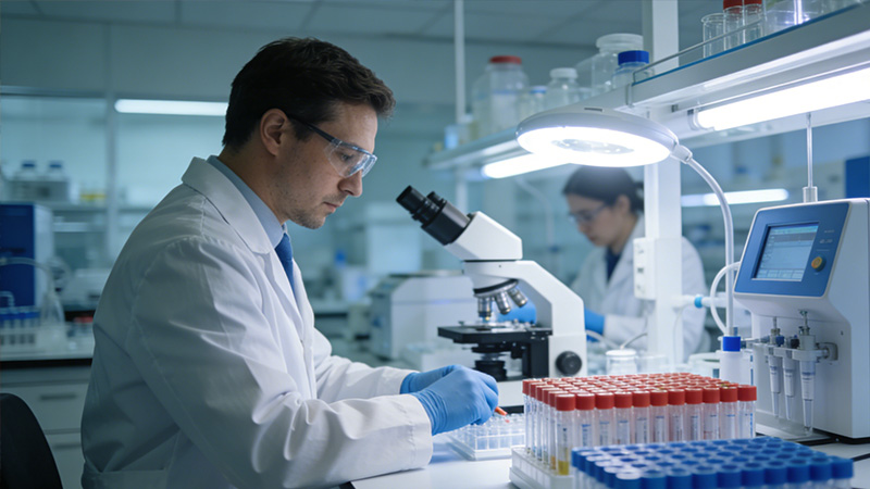
Medical imaging solutions
A new shape for medical imaging: from manual reading to AI-assisted diagnosis
Imaging volume keeps growing while reading capacity is unevenly distributed. Traditional reading workflows hit three pain points: low throughput, large inter-reader variability, and missed subtle findings. Deep learning and multimodal models are now becoming a real lever for accuracy and for spreading specialist knowledge beyond top hospitals.
Four AI engines for precision medicine
1. Multimodal imaging analysis
A leading unstructured-data pipeline reads CT, MRI, X-ray, pathology slides, and ultrasound together, then performs joint detection, segmentation, and quantification. Clinicians get precise second-opinion support on lesion detection, boundary, and severity.
2. Knowledge-augmented clinical decision
RAG binds medical textbooks, clinical guidelines, recent literature, and historical cases into a specialty knowledge base. As the model analyzes an image, it not only marks lesions but also proposes differential diagnoses, cites references, and surfaces similar cases, moving from "see" to "read."
3. Workflow integration
A medical-grade workflow embeds AI analysis into PACS and the diagnostic chain: upload, AI pre-analysis, clinician review, report generation, follow-up. The end-to-end automation removes the manual handoffs that slow departments down.
4. Trustworthy AI and compliance
Enterprise deployment keeps data inside the hospital network or a private cloud. The model produces explainable outputs with visual evidence for each lesion, and multi-layer content checks plus audit logging satisfy medical data security and ethics requirements.
Where it applies across the diagnostic chain
Early screening and lesion detection
- Lung nodule screening. CT nodules at or above 3 mm are detected automatically and stratified for malignancy risk, with sensitivity above 98 percent.
- Stroke triage. In the emergency room, CT images are read for hemorrhage and infarction in under one minute, supporting thrombolysis decisions.
- Breast microcalcification. Tiny calcifications on mammography are surfaced reliably, lifting early screening accuracy.
Quantification and assessment
- Liver fat quantification. MRI-PDFF delivers non-invasive liver fat measurement, replacing biopsy in NAFLD staging.
- Surgical planning. Cobb angle, joint replacement angles, and other key parameters are measured automatically for precise digital planning.
- Treatment response. RECIST 1.1-aligned comparison of pre- and post-treatment lesions generates quantitative response reports.
Reporting and knowledge
- Structured report drafts. AI findings seed a structured draft that the clinician confirms or edits, saving 70 percent of report time.
- Cross-modal correlation. Imaging findings are linked to history and labs, flagging potential contradictions or supporting evidence.
- Teaching and quality control. Difficult and typical cases are tagged automatically, building a teaching library and supporting consistency audits.
Clinical impact: efficiency, quality, and consistency
| Dimension | Traditional manual reading | AI-assisted diagnosis |
|---|---|---|
| Reading throughput | Per-clinician daily ceiling | AI pre-analysis lifts throughput 3 to 5x |
| Sensitivity | Fatigue and experience dependent, subtle lesions missed | Stable 95%+ sensitivity on key lesions |
| Report standardization | Variable phrasing, key fields often missing | Structured output with full, consistent fields |
| Clinician training | Long experience-driven path | AI as a second set of eyes for junior clinicians |
| Resource access | Specialist capacity concentrated in major cities | Cloud or appliance deployment extends reach to primary care |
Toward an intelligent, integrated future of diagnosis and treatment
The value of medical AI is not just reading images; it is pulling imaging into the full clinical workflow. With leading tertiary hospitals, we are moving from assistant tool to decision participant, exploring AI-driven prognosis, treatment recommendation, and personalized follow-up.
Hospitals, imaging centers, and research teams: reach out to evaluate the platform and discuss collaboration scenarios.